What Is Histopathology?
Histopathology is the study of tissue or cells to identify signs of disease and injury. It can also detect pathogens such as bacteria, parasites and fungi. It uses tissue samples obtained through procedures like colonoscopies, colposcopies, endoscopies and breast biopsies.
Histology is the study of tissues, while pathology studies disease, hence the name. A histopathologist examines human tissue under a microscope to look for cancer features or other abnormalities. They then make a diagnosis based on their discoveries and compile their findings into a pathology or biopsy report. This report might include:
- A detailed description of the tissue’s appearance
- The diagnosis
- Estimated size of the tumor and its margins (if applicable)
- A synopsis of the histopathologist’s findings
- Any other relevant comments about the sample
Following the diagnosis, a histopathologist can help the patient’s primary doctor or physician determine a treatment route and manage the patient’s care.
The Tissue Examination Process
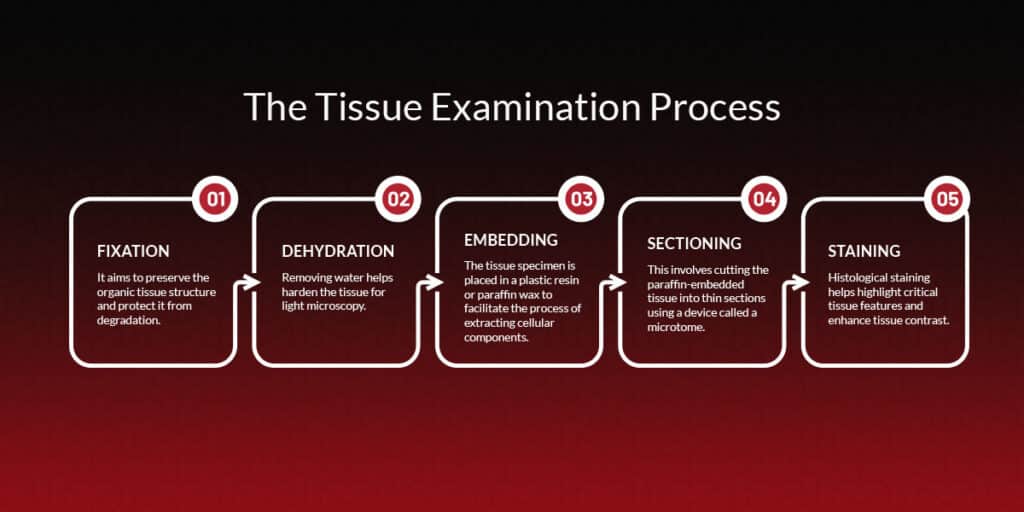
Tissue specimens undergo various steps to prepare them for microscope inspection and disease detection. Here are the five main steps involved with histological tissue examination.
1. Fixation
Fixation is the application of chemicals to irreversibly cross-link proteins. It aims to preserve the organic tissue structure and protect it from degradation. Neutral buffered formalin is typically used for fixation when conducting the investigation under a light microscope. However, other fixatives like glutaraldehyde and formalin can be used.
2. Dehydration
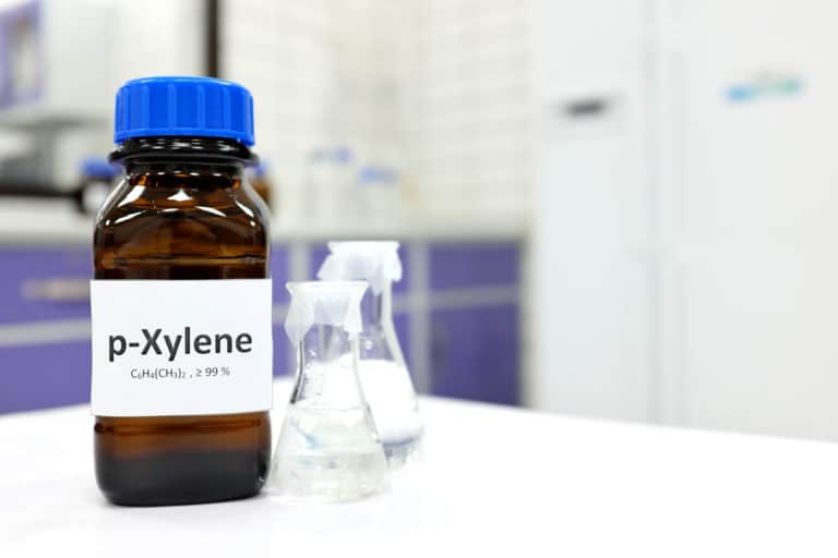
The histologist adds ethanol to dehydrate the sample. Removing water helps harden the tissue for light microscopy. After successful tissue dehydration, the histologist removes the ethanol with xylene.
3. Embedding
During embedding, the histologist puts the tissue specimen into a plastic resin or paraffin wax to facilitate the process of extracting cellular components.
Embedding requires extreme caution and care. Prolonged heating from these fixatives can break down the cell and tissue structures, causing issues with hybridization. Additionally, paraffin wax can impede the penetration of antibodies, chemicals and fixatives, potentially causing a false result.
4. Sectioning
This step involves cutting the paraffin-embedded tissue into thin sections using a device called a microtome. It can then be mounted below the microscope for investigation.
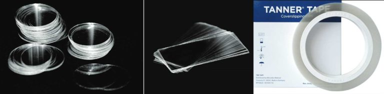
To allow light to pass through the sample and enhance visibility, the ideal thickness for each section is 4-5 micrometers. The histologist must cover the specimen with a coverslip film or cover glass before proceeding with microscope investigation. Sectioning is a crucial process for obtaining accurate details and results and determining the tissue’s abnormality.
5. Staining
Histopathologists often use chemical stains to interrogate tissue samples. Histological staining helps highlight critical tissue features and enhance tissue contrast. Coloring otherwise transparent tissue sections lets histopathologists view bacteria and abnormal tissue structures more easily.
Hematoxylin is the most common staining dye in histology and pathology. It typically gives the nuclei a deep blue-purple color. It’s often used with the dye eosin, which gives the cell’s nucleus a bright pink hue.
Histology vs. Histopathology
What is histology, and how does it differ from histopathology? Histology encompasses the study of tissues and their microscopic structure. Essentially, histopathology is a branch of histology. It specifically examines diseased and damaged tissue.
Why Are Histopathological Examinations Important?
Histopathologists play an important role in medical diagnoses. They assist doctors and clinicians by performing biopsies — examinations of small tissue samples from the skin, kidney, liver or another organ.
They can determine the nature and extent of the abnormality. If they discover a malignant tumor, they can inform the clinician about the type and grade of cancer, along with its responsiveness to different treatments.
Histopathology can diagnose the following conditions and others:
- Cancers
- Crohn’s disease
- Infections
- Ulcerative colitis
- Uterine fibroids
By inspecting the specimen carefully under a microscope, the histopathologist can identify changes in cell structure that may explain a patient’s disease. The sooner a condition is diagnosed, the sooner the patient can receive the treatment they need. That’s what makes histopathology so essential to the health care field.
What Equipment Is Needed for Histopathology?
Histopathologists need high-quality equipment to obtain detailed, accurate tissue samples. Histology equipment can also automate tedious and repetitive processes, allowing you to focus on more important tasks.
1. Microtomes
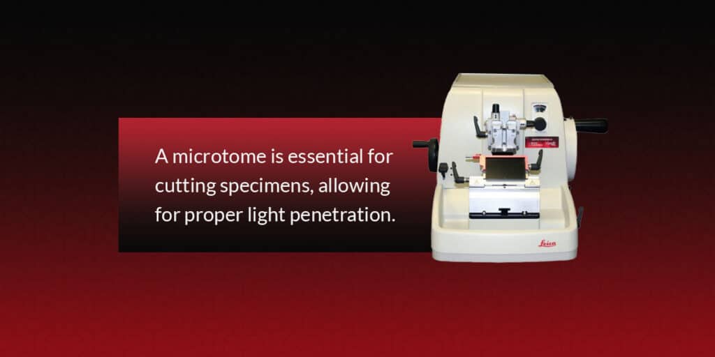
As discussed above, the sectioning process involves slicing the sample into thin sections. A microtome is essential for cutting specimens, allowing for proper light penetration.
Whether you opt for a new or refurbished model, this device offers a quick, simple alternative to manual cutting methods, getting you results faster. You can also choose from multiple configurations, including automated, rotary and sliding. We also recommend microtome lubricant to reduce friction and enable easier movement of the components.
2. Coverslippers
A coverslip must be placed over the sample before microscope examination. A coverslipper is a machine that automatically lays a coverslip film or glass cover over a microscope slide. The film or glass sheet protects the specimen from damage. This convenient technique prevents the need to lay coverslips manually, allowing you to focus on other tasks.
3. Slide Stainers
A slide stainer automatically applies colored dye, peroxidase, antibodies and conjugated enzymes to a tissue sample. The stain penetrates certain cell components, increasing the contrast between different parts.
This process makes microscope examination much simpler by highlighting key areas. You also don’t have to worry about staining by hand, which can be both costly and time-consuming.
4. Embedding Stations
An embedding station helps embed paraffin wax into tissue specimens. Paraffin supports the tissue and retains its shape during sectioning, making the process much easier.
Since an embedding station manages paraffin embedding, you don’t have to complete this process manually and can handle other tasks in the meantime. The machine can also embed tissues more efficiently, streamlining your workflow immensely.
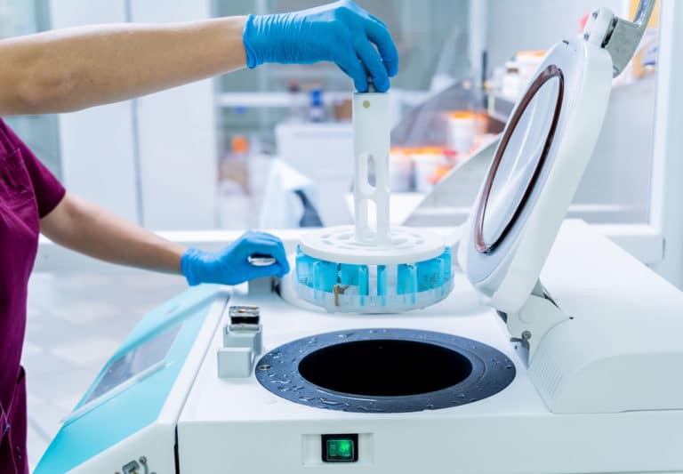
5. Tissue Processors
Cutting a tissue sample without proper preparation can distort or damage its structure. A tissue processor machine infuses the specimen with liquids or paraffin wax, preparing the sample for further analysis.
This solution helps keep the specimen in place during sectioning. A tissue processor can produce higher sample quantities faster than manual methods. It can also yield better-quality sections.
Find Histopathology Equipment From Our Selection Today
At Histologyequipment.com, we have a wide variety of new laboratory equipment and refurbished laboratory equipment. Whether you’re looking for microtomes, tissue processors, slide stainers or other devices, we can work with you to find affordable solutions for your needs. Our products allow you to enjoy more efficient processes and cut down on manual labor.
Choose Histologyequipment.com for reliable and cost-effective histopathology equipment. Browse our product selection or contact us with questions about finding the right products today.