Progressive and Regressive Staining
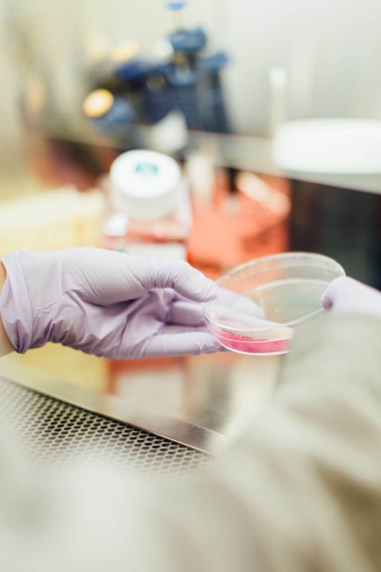
Staining is a special process that differentiates tissue components, allowing for easy observation under a microscope. Hematoxylin and Eosin (H&E) staining is the standard tissue staining procedure in the histology field. A process used consistently in histopathology labs, H&E staining gives histologists and pathologists a highly detailed view of the tissue samples they are examining.
When cell structures such as cytoplasm, nucleus, organelles and extracellular components are precisely stained, the enhanced contrast provides better differentiation of a tissue’s various cellular compartments. Read on to learn more about the different types of H&E staining methods and how they differ from one another.
H&E Staining — The Most Popular Staining Process in Histology and Histopathology Labs
Staining tissues with various dyes to heighten the contrast of the microscopic images is a standard histological process. H&E staining procedures utilize two separate dyes — Hematoxylin and Eosin. This stain combination enhances tissue view, as each dye highlights different elements of the tissue being examined and brings to the forefront the biochemical differences of a tissue sample. Here are the main differences between these stains:
- Hematoxylin: This positively charged dye features the reaction of a basic dye and appears as a purple-to-blue stain color on a tissue sample. Hematoxylin stain adheres to the DNA and nucleoprotein-holding cell nucleus and also highlights the RNA-containing organelles, including ribosomes and the endoplasmic reticulum.
- Eosin: This more acidic and negatively charged dye typically stains tissue components in a red-to-pink color. Eosin stain adheres to basic tissue structures, including the cell walls, cytoplasm and extracellular fibers.
By using the proper H&E staining method, histologists produce important views of normal and abnormal cell and tissue characteristics.
Progressive, Modified Progressive and Regressive H&E Staining Methods
H&E staining processes are classified into three different types — progressive, regressive and modified progressive.
Progressive Staining
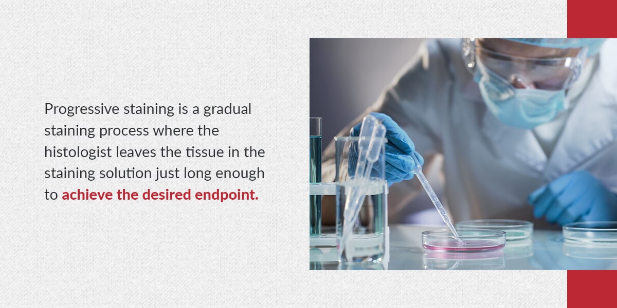
Progressive staining is a gradual staining process where the histologist leaves the tissue in the staining solution just long enough to achieve the desired endpoint. It uses a weaker H&E solution than regressive staining. Progressive stains allow for a higher result intensity due to a longer staining process.
The histologist must frequently monitor the stain quality to deem the staining process complete. The immersing time influences the staining intensity. The histologist must stop the process at just the right time to achieve the stain’s planned-upon intensity.
Unlike regressive staining, progressive staining does not require differentiation (excess stain removal). Progressive stains have a low Hematoxylin concentration, while regressive stains have a high Hematoxylin concentration. Therefore, progressive stains slowly and carefully stain the chromatin.
Mayer’s and Gill’s Hematoxylins are naturally progressive, making them typical dyes for progressive staining. Histologists often use them as a nuclear counterstain for immunohistochemistry purposes and special stains. A staining time of about 5-10 minutes is standard for these two Hematoxylins. Harris Hematoxylin is another commonly used Hematoxylin, particularly for progressive staining cytology specimens.
Progressive staining highlights mucin and other noncellular tissue material by the Hematoxylin. Any staining of extracellular tissue components can help better indicate tumors that are well-differentiated.
Regressive Staining
Regressive staining is a quicker staining process than progressive staining. The histologist deliberately over-stains the tissue until all of its components are dye-saturated. Then, they de-stain the tissue until it reaches the desired endpoint. As mentioned previously, this de-staining step is called differentiation.
Most of the time, de-staining is achieved using an acidic solution made up of dilute HCL, or acetic acid in alcohol or in water. After de-staining, the slide is “blued” by rinsing the lens under tap water until blue sections appear on the sample.
When very clear differentiation of tissue elements is required, histologists prefer regressive staining because of the customized stain intensity that results from the process. Because regressive stains have a high Hematoxylin concentration, they quickly diffuse the entire cell, overstaining the cytoplasm and chromatin. Harris Hematoxylin is a commonly used Hematoxylin for regressive staining, but Delafield’s, Ehrlich’s and Mayer’s Hematoxylins can also be used.
Despite the differences between these two procedures, both progressive and regressive staining techniques can accomplish nuclear staining with Hematoxylin.
Modified Progressive Staining
Rather than removing excess stain from the nuclei, modified progressive staining uses a mild differentiator to remove background staining. What makes a stain progressive is the presence of an altered nuclear stain — not a differentiator. Similar to progressive staining, the differentiator should not be too strong. Otherwise, the nuclei will be stained too pale to read properly.
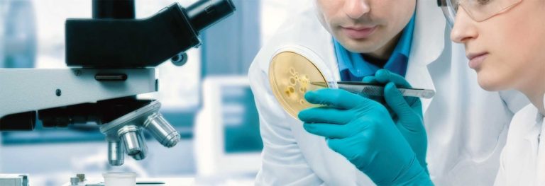
How Can a Slide Stainer Help the Staining Process?
A slide stainer automatically applies colored dye, antibodies, peroxidase and conjugated enzymes to a tissue specimen. The stain penetrates different cell elements, increasing the contrast between specimen parts. A stained slide highlights areas of interest in the tissue sample. That way, you can better examine and assess these regions under a microscope.
Without a slide stainer, you’ll need to apply the stain manually. Staining by hand is more costly and time-consuming. A slide stainer helps improve your throughput and precision. An automated stainer is ideal for histological, biological, cytological and hematologic smearing procedures.
If you’re seeking a new or refurbished automated stainer for your histology lab, look no further than Histologyequipment.com. Our extensive selection includes various options for different specifications and procedures. Regardless of the slide stainer you choose, you’ll receive a reliable, durable device at a highly affordable price.

View Our Collection of Manual and Automated Stainers Now
The highly clarified stains you need require high-quality, accurate stainers. Luckily, you can find the stainers you need at Histologyequipment.com. Choose from our full inventory of new and refurbished slide stainers, as well as stainers fully refurbished by our certified in-house technicians. And go ahead and discover our range of superior Hematoxylin Stains.
Accelerate and simplify the staining process with a quality slide stainer from Histologyequipment.com. Contact us today with any questions about your histological laboratory needs.